The Nerve Fibers in the Dermis Stimulate
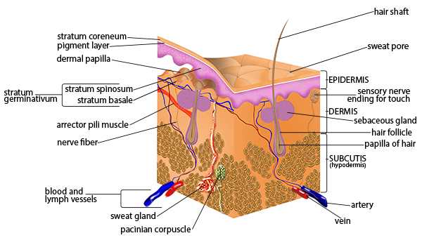
Nerve fibers in the dermis stimulate Muscles and glands in the dermis Two thieves steal jewelry and then drop it as they are escaping. Myelinated nerve bundles located deep in the dermis travel roughly parallel to the surface of the skin with individual myelinated fibers branching off more superficially in the dermis where they course perpendicular to the skins surface to innervate mechanoreceptors and terminate in dermal papillae Fig.
Seer Training Anatomy Of The Skin
Fat cells in the subcutaneous layer.
. Updated 3132014 101743 PM. They are most likely. Fibers in the dermis are produced by fibroblasts.
The police recover the jewelry and an officer explains on the evening news the fingerprints were obtained from the back of the watch. The nerve fibers in the dermis stimulate A. Muscles and glands in the dermis.
Melanocytes transfer melanin granules to. Sweat glands blood vessels and the arrector pili muscle are innervated by sympathetic C-fibers in the dermis. The ________ layer of the dermis contains bundles of collagen fibers and elastin and is responsible for the mechanical strength and flexibility of the skin.
What are the arrector pili muscles responsible for. The epidermis blood vessels and skin appendages such as hair follicles sebaceous glands sweat glands and apocrine glands are innervated by several subtypes of sensory nerves. Asked Oct 22 2021 in.
D all of the above. Asked Aug 7 2019 in Anatomy Physiology by coxjc. The nerves will let you feel texture and temperature of your environment and the brain will react depending on that stimuli example.
Melanocytes in the epidermis. The nerve fibers in the dermis stimulate the muscles and the glands in the dermis. Two thieves steal jewelry and then drop it as they are escaping.
B muscles and glands in the dermis. If you touched a very hot surface your brain will send signals to your muscles to make you move your hands and arms away from that harmful stimuli. A muscles and glands in the dermis.
Both SP and NKA have been shown to stimulate proliferation of human cultured. The major blood vessels that supply the skin are in the A. The police recover the jewelry and an officer explains on.
The nerve fibers in the dermis stimulate answer choices muscles and glands in the dermis. You step out of the shower and vigorously rub your skin with a towel. Muscles and glands in the dermis.
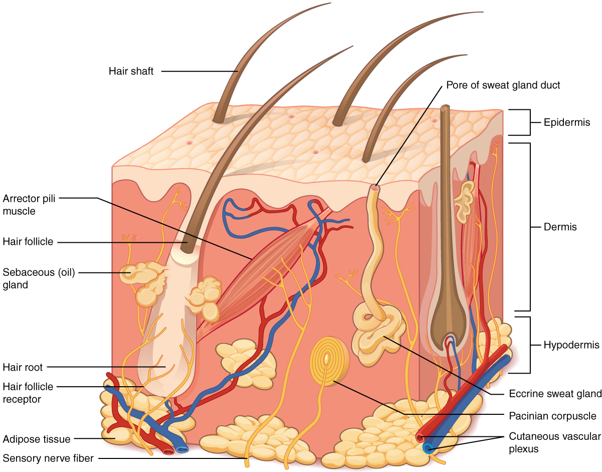
Fat cells in the subcutaneous layer. The major blood vessels that supply the skin are in the. Those that are continuous with the dermis and are intertwined with dendrites are called Free nerve endings and tiny blood vessels are characteristic of the papillary layer of the dermis.
A tactile receptor is composed of a capsule that surrounds a core of collagen fibers. Afferent intraepidermal nerve fibers of the class C and Aδ are found in the epidermis as free nerve endings. Most of the nerve fibers are found in the mid-dermis and the papillary dermis.
Asked 3132014 95728 PM. Asked Nov 13 2016 in Health Professions by FabKid. C melanocytes in the epidermis.
Epidermis As cells are pushed from the deeper portion of the epidermis toward the surface They die The dermis is composed largely of Dense irregular connective tissue Nerve fiber scattered throughout the dermis are associated with Muscles glands and sensory receptors. Answered Nov 13 2016 by Emir02. Expert answeredkclementiPoints 0 User.
Blood vessels in the epidermis. The skin is innervated by afferent somatic nerves with fine unmyelinated c or myelinated aδ primary afferent nerve fibers transmitting sensory stimuli temperature changes chemicals inflammatory mediators ph changes via dorsal root ganglia and the spinal cord to specific areas of the cns resulting in the perception of pain burning. B blood vessels in the epidermis.
The nerves provide the brain stimuli from which it can react from. D fat cells in the subcutaneous layer. The nerve fibers in the dermis stimulate A blood vessels in the epidermis.
Blood vessels in the epidermis. D More questions like this Free nerve endings and tiny blood vessels are characteristic of the papillary layer of the dermis. Log in for more information.
The nerve fibers scattered throughout the dermis are associated with sensory receptors. What are the types of. Melanocytes in the epidermis.
What makes fibers in the dermis. The nerve cells in the dermis sense. Muscles and glands in the dermis.
D fat cells in the subcutaneous layer. If you were able to analyze the towel you would find skin cells. What motor nerves stimulate the arrector pili muscles.
The nerve fibers in the dermis stimulate. Two thieves steal jewelry and then drop it as they are escaping. 75 rows The nerve fibers in the dermis stimulate.
Question 11 30 seconds Q. C melanocytes in the epidermis.

Skin Care Tip Guide How Does Collagen And Elastin Work Subcutaneous Tissue Skin Structure Epidermis
Chapter 5 Integument System
Layers Of The Skin Anatomy And Physiology

A Simplified Illustration Of The General Anatomy Of The Skin With The Download Scientific Diagram
No comments for "The Nerve Fibers in the Dermis Stimulate"
Post a Comment